Fluorescent PMMA Microparticles
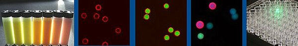
PolyAn's fluorescent microparticles are available in various sizes, emission spectra and fluorescence intensities. The fluorescent PMMA microparticles are suitable for use in flow cytometry, fluorescence microscopy, phagocytosis studies and cell labelling. They can be used in image based systems as well as in other screening applications. Typical applications include calibration of flow cytometers, calibration of fluorescence microscopes and multiplex bead assays.
With PolyAn’s production process up to six fluorophores can be incorporated into the beads during the bead polymerisation process. This ensures a much more homogeneous distribution of the dyes within the beads when compared to conventional diffusion controlled dyeing processes. The fluorophores are also caged within the polymeric PMMA matrix and are less likely to leak-out.
The standard packaging volume for our fluorescent, unmodified bead is 1.5 mL. The beads are available in any size between 2 - 20 µm. Both the fluorescence intensity and the spectral characteristics can be tailored to your specific requirements as part of our Molecular Surface Engineering Service.
Our fluorescence encoded beads can also be functionalized with 3D-Carboxy, Streptavidin, Neutravidin, Protein A/G, 3D-Azide and 3D-Alkyne surfaces. A custom modification with antibodies or oligonucleotides is available upon request. Please do not hesitate to contact us!
Selected Publications and References
- Weber, T.A. et al., `Single-Color Barcoding for Multiplexed Hydrogel Bead-Based Immunoassays´, Appl. Mater. Interfaces, 2022, 14, 25147. DOI: 10.1021/acsami.2c04361.
- Bergmann, S. et al., `NO Synthesis in Immune-Challenged Locust Hemocytes and Potential Signaling to the CNS´, Insects, 2021, 12, 951. DOI: 10.3390/insects12100951.
- Rodríguez-Pena, A. et al., `Spheroscope: A custom-made miniaturized microscope for tracking tumour spheroids in microfluidic devices´, Sci. Rep., 2020, 10, 2779. DOI: 10.1038/s41598-020-59673.
- Ahlfeld, T. et al., `Bioprinting of mineralized constructs utilizing multichannel plotting of a self-setting calcium phosphate cement and a cell-laden bioink´, Biofabrication, 2018, 10, 45002. DOI: 10.1088/1758-5090/aad36d.