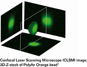
Fluorescent Microparticles
PolyAn offers a wide range of fluorescent PMMA microparticles (beads) in sizes between 2–20 µm for multiplex bead assays, calibration tools, and fluorescence lifetime-based applications.
The fluorescence intensity of the beads can be tailored to specific applications and read-out systems. We are happy to help you select a suitable fluorescence intensity for your application. Customized coupling of biomolecules is available upon request. Please do not hesitate to contact us if you have a specific request!
PolyAn also offers Fluorescent PMMA Nanoparticles in the 100–500 nm size range.

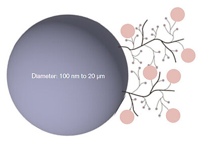
Functionalized Fluorescent Microparticles
PolyAn's fluorescent beads can be equipped with a broad range of functional surfaces:
- Unmodified fluorescent beads or modified with a Low aggregation surface
- Reactive surfaces: 3D-Carboxy and 3D-Aldehyde
- Surfaces for Click chemistry: 3D-Azide, 3D-Alkyne, 3D-DBCO, and 3D-MTZ
- Functionalized with Streptavidin, Neutravidin, or Protein A/G
A custom modification with other functional groups (e.g. Maleimide, Biotin,…), with different loading capacities, and the customised coupling of biomolecules (Oligonucleotides, Peptides, Antibodies …) are also available upon request. Please do not hesitate to contact us!
Fluorescence encoding toolbox
PolyAn’s fluorescent micro- and nanobeads are encoded with either one fluorophore (single-color) or multiple fluorophores (multi-color) at excitation/emission wavelengths and fluorescence intensities of your choice.
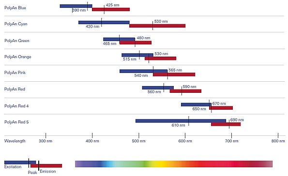
PolyAn beads can be color-encoded with up to six fluorescent dyes. Multiplex beads with specific intensity ratios or fluorescence lifetimes as well transparent and dye-colored (non-fluorescent) beads are also available.
With PolyAn’s production process the fluorophores are incorporated into the beads during the polymerization process. This ensures a much more homogeneous distribution of the dyes within the beads when compared to conventional diffusion controlled dyeing processes. Additionally, the fluorophores are caged within the polymeric matrix and are less likely to leak-out.
Please have a look at our fluorescence encoded products. PolyAn also produces customized microparticles which incorporate fluorophores for other spectral ranges.
Selected Publications and References
- Pham, N.M. et al., `Immuno-gold silver staining assays on capillary-driven microfluidics for the detection of malaria antigens´, Biomed. Microdevices, 2019, 21, 24. DOI: 10.1007/s10544-019-0376.
- Pham, N.M. et al., `A bead-based immunogold-silver staining assay on capillary-driven microfluidics´, Biomed. Microdevices, 2018, 20, 41. DOI: 10.1007/s10544-018-0284-6.
- Schauer, O. et al., `Motility and chemotaxis of bacteria-driven microswimmers fabricated using antigen 43-mediated biotin display´, Sci. Rep., 2018, 8, 9801. DOI: 10.1038/s41598-018-28102-9.
- Mostaghaci, B. et al., `Bioadhesive Bacterial Microswimmers for Targeted Drug Delivery in the Urinary and Gastrointestinal Tracts´, Adv. Sci., 2017, 4, 1700058. DOI: 10.1002/advs.201700058.