Focus Beads for Cell Imaging
PolyAn’s Focus Beads are used to quickly find the correct focal plane via auto-focus in automated imaging system, which is a key challenge for reliable cell-based assays.
The 2 μm Focus Beads have a comparable size to cell nuclei and consist of a biocompatible PMMA grade that is not cell-toxic and does not interfere with cell behavior. The Focus Beads are color-encoded with our PolyAn Blue dye, which emits in the DAPI channel, but is transparent in all other detection channels.
PolyAn Focus Beads
Key features
In most applications DAPI-focusing on cell nuclei is used to determine the bottom of slides, plates, or other substrates. However, this method can lead to errors when the cells detach from the bottom surface or are insufficiently stained.
PolyAn’s Focus Beads, in turn, have a well-defined fluorescence intensity, thus, avoiding problems caused by insufficient staining. The beads can be easily added to the cell suspension and quickly sink to the bottom to indicate the correct focal plane.
Application example
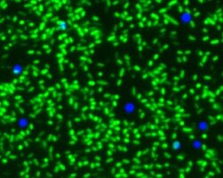
In the examples illustrated below the read-out was done using the VideoScan HCU, a fluorescence imaging system based on a fluorescence microscope.* The microscope focusses quickly on the focus beads independent whether bacteria adhere or not:
Focus beads (blue, DAPI channel) in combination with EPEC-bacteria on a GP2-coated plate. Only the surface of the bacteria has been dyed with O26 E.coli-antibody sera and FITC-conjugated secondary antibody.
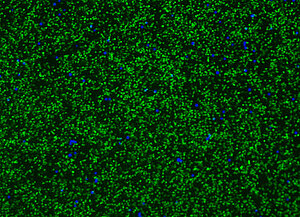
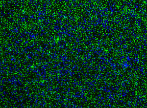
Focus beads (blue, DAPI channel) in combination with E.Coli-bacteria on a GP2-coated plate. The bacteria have been colored using PI (DNA).
Publications & downloads
- Bartlitz, C. et al., `Adhesion of Enteropathogenic, Enterotoxigenic, and Commensal Escherichia coli to the Major Zymogen Granule Membrane Glycoprotein 2´, Appl. Environ. Microbiol., 2022, 88, 02279-21. DOI: 10.1128/aem.02279-21.
- Rödiger, S. et al., `A Highly Versatile Microscope Imaging Technology Platform for the Multiplex Real-Time Detection of Biomolecules and Autoimmune Antibodies´, Adv. Biochem. Eng. Biotechnol., 2013, 133, 35. DOI: 10.1007/10_2011_132.
