Use of focus beads for imaging of cell-based assays
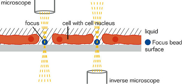
Quickly finding the correct focus level is one of the key challenges for automated, microscope based fluorescence maging systems. In most applications DAPI-focussing on cell nuclei is used to determine the bottom of slides, plates or other carrier materials. However, this method can lead to errors when the cells detach from the bottom surface or are insufficiently stained.
PolyAn addresses this problem with our new 2µm blue Polymethylmethacrylate (PMMA) beads:
- 2 µm beads have a comparable size to cell nuclei.
- PMMA-grade is not cell-toxic and does not interfere with the cell behavior.
- Color encoded in the DAPI channel, transparent in all other channels.
- Well-defined fuorescence intensity. Problems caused by insuffcient staining of the cell nuclei can thus be avoided.
- Easy handling: the beads can be easily added to the cell suspension. They quickly sink to the bottom and thus indicate the correct focus level.
- Application: standards in a range of cell assays, e.g. bioflim assays, adhesion assays or detection of bacteria.
The beads have a well-defined fluorescence intensity. Problems caused by insufficient staining of the cell nuclei can thus be avoided. PolyAn‘s focus beads can be used as standards in a range of cell assays, e.g. biofilm assays, adhesion assays, detection of bacteria.
2 µm, unmodified PMMA microparticles, PolyAn Blue
| Id | 105 89 002 |
| Title | 2 µm, unmodified PMMA microparticles, PolyAn Blue |
| Substrate | PMMA |
| Format | Microparticles |
| Mean Diameter | 2 µm |
| Color Labeling | PolyAn Blue |
| Surface Modifications | unmodified |
| Solids Content | 1% |
| Product Dimensions | |
| Packaging | Aqueous Suspension |
| Packaging Volume | 1.5 mL |
| Package Weight | 5 g |
| Dimensions | 35 x 15 x 15 mm |
| Hts Code | 3906 1000 |
| Pads Wells | |
| Pad Size | |
| Well Format | |
| Product Thickness | |
| Description | |
| Image |
In the examples illustrated below the read-out was done using the VideoScan HCU*, a fluorescence imaging system based on a fluorescence microscope**. The microscope focusses quickly on the focus beads independent whether bacteria adhere or not:
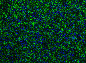
Focus beads (blue, DAPI channel) in combination with EPEC-bacteria on a GP2-coated plate. Only the surface of the bacteria has been dyed with O26 E.coli-antibody sera and FITC-conjugated secondary antibody.
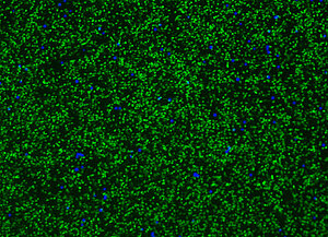
Focus beads (blue, DAPI channel) in combination with E.Coli-bacteria on a GP2-coated plate. The bacteria have been colored using PI (DNA).
Publications & downloads
* Rödiger, S. et.al. Adv. Biochem. Eng. Biotechnol. 2013, 133, 35–74
** Schierack, P. et.al. Gut (BMJ Group), 2014
PolyAn product flyer Focus Beads