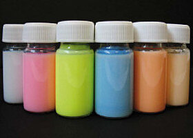
Fluorescent PMMA Nanoparticles
PolyAn’s fluorescent Nanobeads are available in different sizes from 100 nm to 500 nm. They are characterized by a narrow particle size distribution and an excellent batch-to-batch reproducibility. PMMA Nanobeads are non-toxic, and thus, suited for live cell applications.
PMMA Nanobeads are available with various reactive surface functionalizations for immobilization of biomolecules (proteins, antibodies, peptides, oligonucleotides, etc.):
Applications for Nanobeads
We offer a large range of different PMMA Nanobeads suitable for particle-based technologies, including:
- Calibration (e.g. flow cytometry, fluorescence microscopy)
- Lateral Flow Assays
- Solid phase immunoassays
- Immunoturbidimetric assays
- Nephelometric assays
- Cell staining/compartment staining Applications

Size and polymer
All Nanobeads are comprised of polymethyl methacrylate (PMMA). Using PMMA ensures an excellent optical brilliance and low autofluorescence compared to other microparticle materials. The refractive index of 1.48 is close to the refractive index of cells (ca. 1.38). Our microparticles have a density of 1.19 g/cm³ and a glass transition temperature (Tg) of about 110°C. PolyAn uses a biocompatible grade of PMMA.
PMMA Nanobeads are available in distinct, monodisperse populations with (mean) diameters between 100 nm to 500 nm as illustrated below.

DLS measurement of differently sized PMMA Nanobeads.
Please note, that PolyAn also produces customized nanoparticles which incorporate fluorophores for other spectral ranges. All PMMA Nanobeads are available in sizes between 100-500 nm. Custom modifications are available upon request. Please do not hesitate to contact us to get some of our PMMA-Nanobeads!
Selected Publications
- Anyanwu, N.C.J. et al., `Rigorous Process for Isolation of Gut-Derived Extracellular Vesicles (EVs) and the Effect on Latent HIV´, Cells, 2025, 14, 568. DOI: 10.3390/cells14080568.
- Doveri, L.R. et al., `Going hi-res in bulk: flowless multiangle dynamic light scattering for detection on asymmetric flow field flow fractionation.´, J. Mater. Chem. B., 2025, DOI: 10.1039/D4TB02790F.
- Kalita, I. et al., `Motile and Chemotactic Minicells and Minicell-Driven Biohybrids Engineered for Active Cargo Delivery´, Appl. Mater. Interfaces, 2025, 17, 36387. DOI: 10.1021/acsami.5c04638.
- Zhang, Y. et al., `An analytical model of label-free nanoscale chemical imaging reveals avenues toward improved spatial resolution and sensitivity´, PNAS, 2025, 122, 2403079122. DOI: 10.1073/pnas.2403079122.
- Zytowski, E. et al., `Uptake and translocation of nanoplastics in mono and dicot vegetables´, Plant Cell Environ., 2025, 48, 134. DOI: 10.1111/pce.15115.
- Braun, A. et al., `Uptake and Cellular Effects of Polymethylmethacrylate on Human Cell Lines´, Microplastics, 2024, 3, 205. DOI: 10.3390/microplastics3020012.
- Schmidt, C. et al., `Fluorescence-encoded poly(methyl metharcylate) nanoparticles for a lateral flow assay detecting IgM autoantibodies in rheumatoid arthritis´, Anal. Biochem. 2021, 633, 114389. DOI: 10.1016/j.ab.2021.114389.
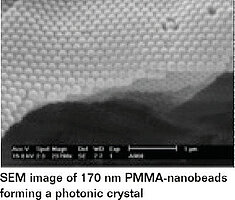
*Image courtesy of BTU Cottbus-Senftenberg