PolyAn Beads for Fluorescence Imaging
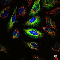
Fluorescence imaging (fluorescence microscopy) is a powerful technique used in biology, medicine, and materials science to visualize and study specific molecules or structures within a sample. It relies on fluorescent dyes or proteins that emit light when excited by a particular wavelength, allowing researchers to detect specific components and observe dynamic processes within cells and tissues with high sensitivity and specificity. This method is widely used in cell biology, molecular biology, and medical diagnostics to observe cellular processes, gene expression, protein localization, and molecular interactions in real time, offering valuable insights into complex biological systems.
Bead-based Calibration Tools
PolyAn’s Multiplex Beads are designed for the development of multiplex assays that can be analyzed using a wide range of flow cytometers or fluorescence imaging-based platforms (e.g. fluorescence microscope systems):
Multiplex Beads for Fluorescence Imaging
PolyAn’s Multiplex Beads are designed for the development of multiplex assays that can be analyzed using a wide range of fluorescence imaging-based platforms (e.g. fluorescence microscope systems):
- Dual color-encoded Multiplex Beads with up to 18 bead populations
- Customized development of multiplex sets for OEM-applications
Selected Publications
PolyAn Fluorescence Lifetime Beads for FLIM:
- Knight, V.R. et al., `Fast wide-field light sheet electro-optic FLIM´, Optics Express, 2026, DOI: 10.1364/OE.581295.
- Kellerer, T. et al., `Speed-Up Phase Resolved Fluorescence Lifetime Imaging Microscopy (SUPER-FLIM) for Real-Time Microenvironmental Sensing´, Res. Square, 2025, DOI: 10.21203/rs.3.rs-6724982/v1.
- Kanno, H. et al., `High-throughput fluorescence lifetime imaging flow cytometry´, Nat. Commun., 2024, 15, 7376. DOI: 10.1038/s41467-024-51125-y.
- Lin, Y. et al., `Coupling a recurrent neural network to SPAD TCSPC systems for real-time fluorescence lifetime imaging´, Sci. Rep., 2024, 14, 3286. DOI: 10.1038/s41598-024-52966-9.
- Kellerer, T. et al., `Rigorous investigation and comparison of different Fluorescence Lifetime Imaging Microscopy (FLIM) techniques analyzed using the phasor plot´, Proc. of SPIE, 2024, 12847, 128470O. DOI: 10.1117/12.3002648.
- Angelone, D. et al., `Time resolved SPAD micro-camera probe for wide-field FLIM in microendoscopy´, Proc. of SPIE, 2023, 12368, 1236807. DOI: 10.1117/12.2648340.
- Nutt, K.J. et al., `High-efficiency digitally scanned light-sheet fluorescence lifetime microscopy (DSLM-FLIM)´, bioRxiv, 2023, DOI: 10.1101/2023.06.02.543377.
PolyAn Beads for Microscopic Assays and Cell Imaging:
- Kalita, I. et al., `Motile and Chemotactic Minicells and Minicell-Driven Biohybrids Engineered for Active Cargo Delivery´, Appl. Mater. Interfaces, 2025, 17, 36387. DOI: 10.1021/acsami.5c04638.
- Kim, J. et al., `Characterization of a Single-Capture Bright-Field and Off-Axis Digital Holographic Microscope for Biological Applications´, Sensors, 2025, 25, 2675. DOI: 10.3390/s25092675.
- Sun, Y. et al., `Engineering Mechanostable Anticalin Scaffolds to Enhance Particle Adhesion and Targeting of CTLA-4 Under Shear Stress´, Angew. Chem., 2025, 64, 4483. DOI: 10.1002/anie.202504483.
- Zytowski, E. et al., `Uptake and translocation of nanoplastics in mono and dicot vegetables´, Plant Cell Environ., 2025, 48, 134. DOI: 10.1111/pce.15115.
- Braun, A. et al., `Uptake and Cellular Effects of Polymethylmethacrylate on Human Cell Lines´, Microplastics, 2024, 3, 205. DOI: 10.3390/microplastics3020012.
- Brock, V.J. et al., `Time-resolved role of P2X4 and P2X7 during CD8+ T cell activation´, Front. Immunol., 2024, 15, 1258119. DOI: 10.3389/fimmu.2024.1258119.
- Picazo-Bueno, J.A. et al., `Off-axis digital lensless holographic microscopy based on spatially multiplexed interferometry´, J. Biomed. Optics, 2024, 29, 22715. DOI: 10.1117/1.JBO.29.S2.S22715.
- Alpsoy; L. et al., `Particle ID: A Multiplexed Hydrogel Bead Platform for Biomedical Applications´, Appl. Mater. Interfaces, 2023, 15, 55346. DOI: 10.1021/acsami.3c12122.
- Bergmann, S. et al., `NO Synthesis in Immune-Challenged Locust Hemocytes and Potential Signaling to the CNS´, Insects, 2021, 12, 951. DOI: 10.3390/insects12100951.
- Rodríguez-Pena, A. et al., `Spheroscope: A custom-made miniaturized microscope for tracking tumour spheroids in microfluidic devices´, Sci. Rep., 2020, 10, 2779. DOI: 10.1038/s41598-020-59673.
- Ahlfeld, T. et al., `Bioprinting of mineralized constructs utilizing multichannel plotting of a self-setting calcium phosphate cement and a cell-laden bioink´, Biofabrication, 2018, 10, 45002. DOI: 10.1088/1758-5090/aad36d.
- Zhuang, J. et al., `Propulsion and Chemotaxis in Bacteria-Driven Microswimmers´, Adv. Sci., 2017, 4, 1700109. DOI: 10.1002/advs.201700109.
PolyAn Dual-color Multiplex Beads for automated Multiplexed Imaging Assays:
- Atiyas, Y. et al., `Combining time domain modulation optofluidics and high dynamic range imaging for multiplexed, high throughput digital droplet assays´, Microsys. Nanoeng., 2025, 11, 93. DOI: 10.1038/s41378-025-00918-2.
- Geithe, C. et al., `A multiplex microchamber diffusion assay for the antibody-based detection of microRNAs on randomly ordered microbeads´, Biosens. Bioelectron., 2024, 18, 100484. DOI: 10.1016/j.biosx.2024.100484.
- Dinter, F. et al., `Immobilisation of Lipophilic and Amphiphilic Biomarker on Hydrophobic Microbeads´, bioRxiv, 2023, DOI: 10.1101/2023.01.10.523433.
- Schmidt, C. et al., `A multiparametric fluorescence assay for screening aptamer–protein interactions based on microbeads´, Sci. Rep., 2022, 12, 2961. DOI: 10.1038/s41598-022-06817-0.
- Nawaz, S. et al., `Rapid Detection of Biofilm Formation by Zoonotic Serovars of Salmonella enterica and Avian Pathogenic E. coli Isolates from Poultry´, Pak. Vet. J., 2020, 40, 527. DOI: 10.29261/pakvetj/2020.066.
- Schmidt, C. et al., `Streptavidin Homologues for Applications on Solid Surfaces at High Temperatures´, Langmuir, 2020, 36, 628. DOI: 10.1021/acs.langmuir.9b02339.
- Choi, Y. et al., `A new reporter design based on DNA origami nanostructures for quantification of short oligonucleotides using microbeads´, Sci. Rep., 2019, 9, 4769. DOI: 10.1038/s41598-019-41136-x.
- Dinter, F. et al., `Simultaneous detection and quantification of DNA and protein biomarkers in spectrum of cardiovascular diseases in a microfluidic microbead chip´, Anal. Bioanal. Chem., 2019, 411, 7725. DOI: 10.1007/s00216-019-02199-x.
- Herrmann, A. et al., `Spatial Separation of Microbeads into Detection Levels by a Bioorthogonal Porous Hydrogel for Size-Selective Analysis and Increased Multiplexity´, Anal. Chem., 2019, 91, 8484. DOI: 10.1021/acs.analchem.9b01586.
- Olowe, O.A. et al., `Phylogenetic grouping and biofilm formation of multidrug resistant Escherichia coli isolates from humans, animals and food products in South-West Nigeria´, Sci. African, 2019, 6, 158. DOI: 10.1016/j.sciaf.2019.e00158.
- Liebsch, C. et al., `Solid-phase microbead array for multiplex O-serotyping of Escherichia coli´, Microchim. Acta, 2017, 184, 1405. DOI: 10.1007/s00604-017-2088-4.
- Schiebel, J. et al., `Genotypic and Phenotypic Characteristics Associated with Biofilm Formation by Human Clinical Escherichia coli Isolates of Different Pathotypes´, Appl. Environ. Microbiol., 2017, 83, 166017. DOI: 10.1128/AEM.01660-17.
- Schmidt, C. et al., `Multiplex localization of sequential peptide epitopes by use of a planar microbead chip´, Anal. Chim. Acta, 2016, 908, 150. DOI: 10.1016/j.aca.2015.12.030.
- Rödiger, S. et al., `Nucleic acid detection based on the use of microbeads: a review´, Microchim. Acta, 2014, 181, 1151. DOI: 10.1007/s00604-014-1243-4.
- Frömmel, U. et al., `Adhesion of Human and Animal Escherichia coli Strains in Association with Their Virulence-Associated Genes and Phylogenetic Origins´, Appl. Environ. Microbiol., 2013, 79, 5814. DOI: 10.1128/AEM.01384-13.
- Rödiger, S. et al., `A Highly Versatile Microscope Imaging Technology Platform for the Multiplex Real-Time Detection of Biomolecules and Autoimmune Antibodies´, Adv. Biochem. Eng. Biotechnol., 2013, 133, 35. DOI: 10.1007/10_2011_132.
- Rödiger, S. et al., `Mikropartikelsysteme für die Nukleinsäurediagnostik´, BioSpectrum, 2013, 19, 153. DOI: 10.1007/s12268-013-0287.
- Frömmel, U. et al., `Multiplex-PCR-Mikropartikel-Assay zum Nachweis bakterieller Gene´, Multiparameteranalytik in Forschung und Praxis, 2011, ISBN: 978-3-89967-703-4. DOI: 10.13140/2.1.4032.6084.
- Grossmann, K. et al., `Multiplex Assessment of Non-Organ-Specific Autoantibodies with a Novel Microbead-Based Immunoassay´, Cytometry A, 2011, 79, 118. DOI: 10.1002/cyto.a.21009.